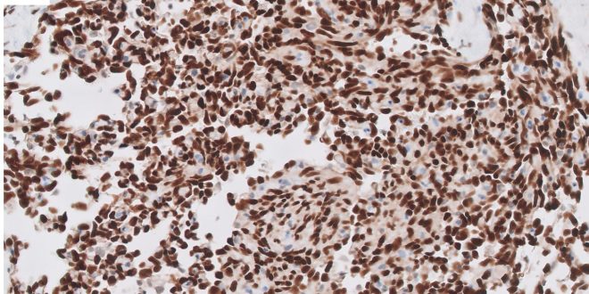
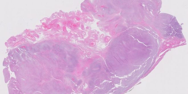
45 year old female with a history of a left lateral thigh mass. Ultrasound reveal a large heterogeneous mixed echogenic solid cystic hypoechoic lesion anterior to the left hip joint likely represents a large complex collection/hematoma. Possibility of a neoplastic lesion cannot be entirely excluded.
Read More »
 Maryland & DC Society of Pathologists Fostering Connections, Inspiring Pathology Excellence
